
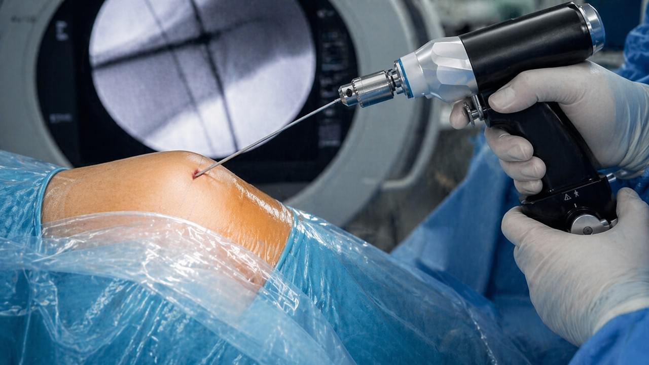
이 글은 당사의 종합 가이드의 일부입니다: 정형외과 전동 공구: 수술용 드릴, 톱 및 시스템 완벽 가이드
요약: 캐뉼라 드릴은 미리 배치된 가이드 와이어 위로 미끄러져 들어가 광범위한 수술 노출 없이 정밀하고 최소 침습적인 골 드릴링을 가능하게 하는 속이 빈 골 드릴입니다. 이 글에서는 캐뉼라 드릴의 기술적 설계, 단계별 MIS 드릴링 기법, 골절 고정 및 인대 재건술에서의 임상적 이점, 그리고 수술팀과 구매 담당자를 위한 주요 선택 기준을 다룹니다.
캐뉼라 드릴이란 무엇인가요?
정의: 캐뉼라 드릴(속이 빈 드릴 또는 가이드 와이어 드릴이라고도 함)은 중앙에 세로 방향의 구멍이 특징인 수술용 골 드릴로, 이미 목표 뼈에 배치된 K-와이어 또는 가이드 핀 위로 드릴이 동축으로 통과할 수 있도록 합니다. 이러한 설계는 최소 침습 수술 (MIS) 정형외과에서 필수적입니다.
“캐뉼라”라는 용어는 라틴어 캐뉼라 (작은 튜브)에서 유래했습니다. 이는 드릴 단면의 속이 비어있는 튜브형 기하학적 구조를 반영합니다. 캐뉼라 드릴은 표준 정형외과용 크기(일반적으로 외경 2.0mm ~ 12.0mm)로 제조되며, 중앙 구멍 직경은 표준 가이드 와이어 크기(1.2mm, 1.6mm, 2.0mm, 2.4mm 및 그 이상)에 맞춰집니다.
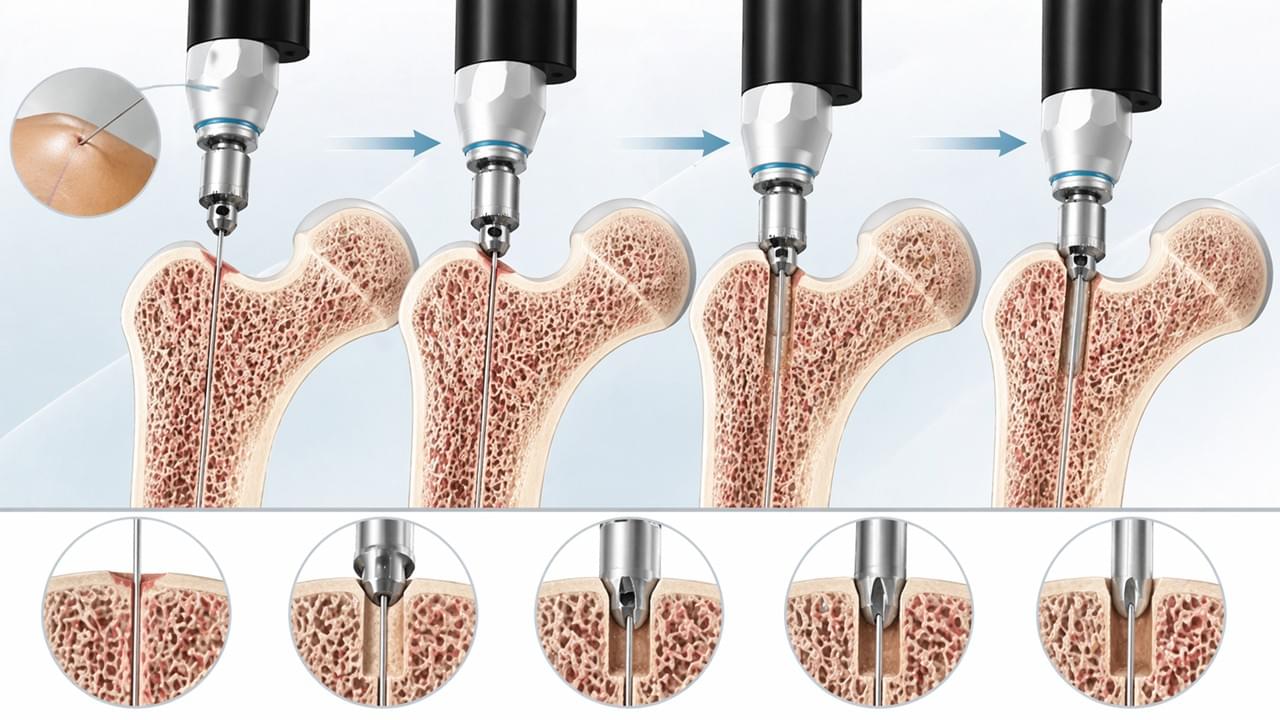
🔧캐뉼라 드릴링 작동 방식: MIS 기법
1단계: 가이드 와이어 배치
형광투시(X-선) 유도 하에 외과의는 작은 절개창을 통해 가이드 와이어(K-와이어 또는 나사형 가이드 핀)를 경피적으로 삽입하고 뼈 내의 정확한 목표 위치로 전진시킵니다. 형광투시는 두 평면에서 정확한 위치를 확인합니다.
이 단계는 중요한 정밀 단계입니다. 가이드 와이어가 나사 또는 임플란트의 최종 경로를 정의합니다.
2단계: 캐뉼라 드릴 삽입
캐뉼라 드릴은 가이드 와이어 위로 끼워지고 동일한 작은 절개창을 통해 뼈 표면으로 전진됩니다. 드릴은 가이드 와이어에 의해 설정된 정확한 경로를 따르며 — 이는 골절 부위를 개방하지 않고도 결과적인 드릴 구멍이 정확하게 위치하도록 보장합니다.
3단계: 동력 드릴링
드릴은 제어된 속도(일반적으로 피질골의 경우 400–1,200 RPM)로 뼈를 통해 전진되며, 열 발생을 관리하기 위해 간헐적인 세척이 이루어집니다. 가이드 와이어는 드릴링 내내 제자리에 유지되어 경로를 유지합니다.
4단계: 가이드 와이어 유지, 나사 삽입
드릴링 후, 캐뉼라 드릴이 제거되는 동안 가이드 와이어는 제자리에 유지됩니다. 그런 다음 캐뉼라 나사가 동일한 가이드 와이어 위로 끼워지고 드릴 구멍으로 삽입되어 — 고정 주기를 완료합니다.
💡 MIS에서 캐뉼라 드릴링의 임상적 이점
| 이점 | 임상적 영향 |
| 최소 절개 | 5–8mm 절개창 대비 5–8cm 개방 접근법 |
| 연조직 보존 | 근육 및 골막 박리 없음; 혈액 공급 유지 |
| 정밀한 경로 | 가이드 와이어가 드릴 경로를 고정; 경로 이탈 없음 |
| 출혈 감소 | 최소한의 연조직 손상으로 수술 중 출혈 감소 |
| 빠른 회복 | 적은 조직 외상 = 적은 통증, 빠른 재활 |
| 낮은 감염 위험 | 작은 상처 표면으로 오염 노출 감소 |
| 형광투시 확인 | 각 단계를 X-ray 하에 확인 후 확정 |
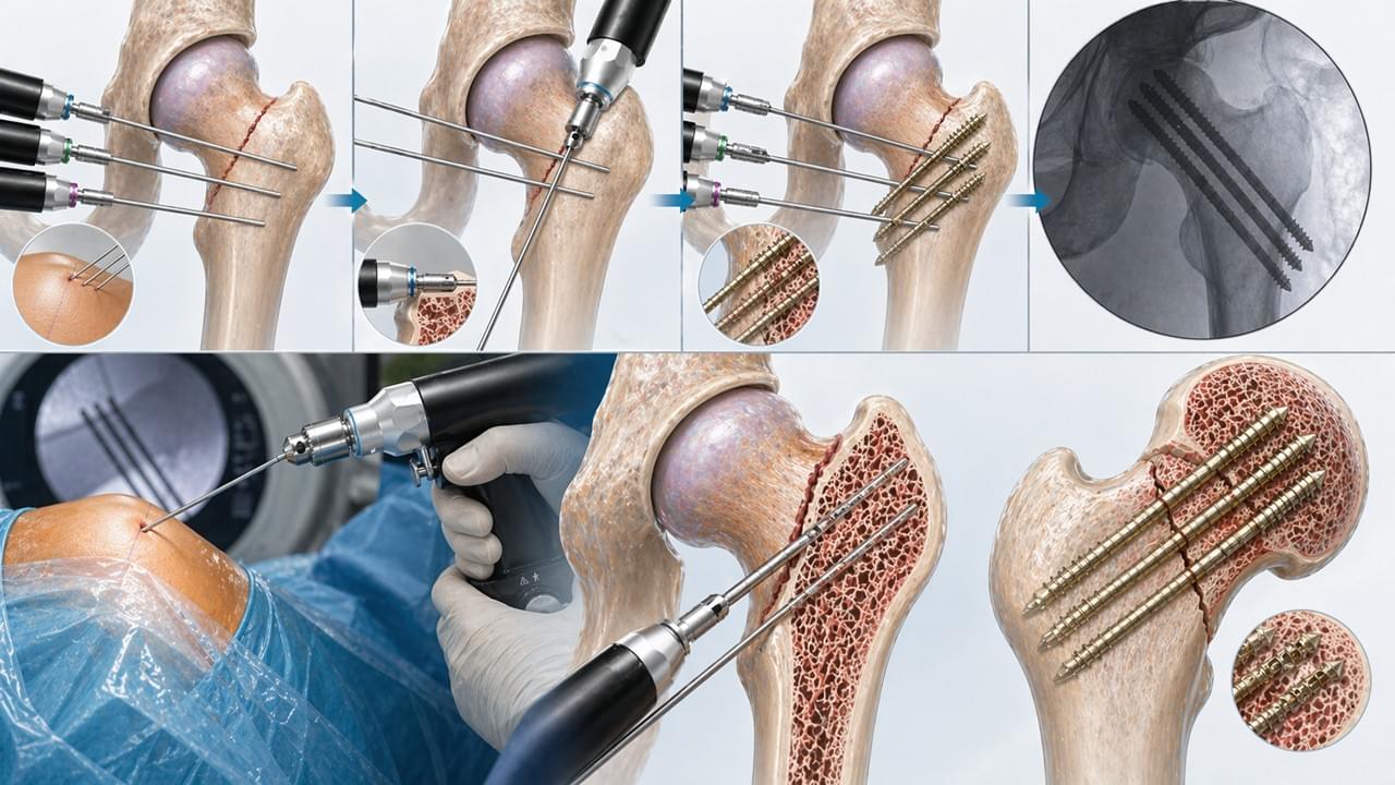
🏥캐뉼라 드릴의 임상 적용
고관절 골절 고정술 (대퇴경부 및 전자간 골절)
가이드 와이어를 통해 삽입되는 캐뉼라 나사는 대퇴경부 골절의 표준 치료법이며, 특히 관절 성형술보다 골절 정복 및 고정을 선호하는 젊은 환자에게 적용됩니다. 세 개의 평행한 캐뉼라 나사는 대퇴골두 혈액 공급을 보존하면서 대퇴골두의 안정적인 고정을 제공합니다.
전방 십자 인대 (ACL) 재건술
관절경적 ACL 재건술은 이식편 통과를 위해 경골과 대퇴골에 골 터널을 생성하는 데 캐뉼라 드릴링 시스템을 사용합니다. 정밀한 터널 배치(해부학적 발자국 정확도)는 이식편 기능에 중요하며, 가이드 와이어 유도 캐뉼라 드릴링을 통해 가능합니다.
주상골 골절 고정술
단일 캐뉼라 무두 압박 나사(Herbert screw)를 이용한 주상골 손목 골절의 경피적 고정은 활동적인 환자에게 황금 표준이 되었습니다. 작고 중앙에 위치한 수근골은 나사 경로에 있어 극도의 정밀성을 요구하며, 이는 캐뉼라 드릴링 기술로만 달성 가능합니다.
소아 골절 고정술 (ESIN/유연성 못)
소아의 탄성 안정성 골수강내 고정술(ESIN)은 캐뉼라 드릴 시스템을 사용하여 대퇴골 및 경골 골절에서 유연한 티타늄 못을 위한 정밀한 피질 진입점을 생성하여 성장판 및 피질 손상을 최소화합니다.
⚙️ 주요 기술 매개변수: 캐뉼라 드릴 시스템 선택
정형외과 또는 수술 키트를 위해 캐뉼라 드릴을 지정할 때, 다음을 평가하십시오:
| 매개변수 | 사양 참고 사항 |
| 외경 범위 | 나사 시스템과의 일치 (예: 4.0mm 캐뉼라 나사는 호환되는 드릴을 필요로 합니다) |
| 중앙 내경 | 최소한의 간격으로 가이드 와이어 직경과 일치해야 합니다 |
| 플루트 형상 | 날카로운 플루트, 골 재료에 적합한 헬릭스 각도 |
| 길이 | 깊은 드릴링을 위해 표준 및 특수 길이로 제공 |
| 재질 | 수술용 스테인리스 스틸 또는 티타늄; 오토클레이브 호환 |
| 연결 방식 | 호환되는 전동 드릴에 퀵 릴리스 또는 AO 연결 방식 |
| 드릴 시스템 호환성 | 기존 전동 드릴 플랫폼과의 호환성을 확인하십시오 |
👉 전체 시스템을 여기에서 살펴보십시오:
🔗 정형외과 전동 공구 제품 페이지
⚠️전문가 자문: 일반적인 캐뉼라 드릴링 오류 방지
Vsun Medical 수술 공학팀:
- 가이드 와이어 굽힘:구부러진 가이드 와이어는 드릴 경로 편차를 유발합니다. 사용 전에 항상 가이드 와이어를 검사하십시오; 어떤 굽힘이라도 보이면 교체하십시오.
- 열 손상:이물질 제거를 위한 빈번한 철수와 함께 간헐적인 드릴링은 드릴 채널의 열 축적을 방지합니다. 지속적인 세척은 열로 인한 골 괴사 위험을 줄입니다.
- 드릴링 중 가이드 와이어 전진:드릴 회전은 가이드 와이어를 뼈 속으로 더 깊이 전진시킬 수 있습니다. 가이드 와이어 홀더를 사용하거나 드릴링 후 투시 하에 와이어 위치를 확인하십시오.
- 드릴 비트 무뎌짐:캐뉼라 드릴 비트는 단면 절삭 면적 감소로 인해 솔리드 비트보다 더 빨리 무뎌집니다. 일회용 비트는 이 위험을 제거하며, 재사용 가능한 비트는 날카로움 확인이 필요합니다.
📚 모범 사례는 다음을 통해 지원됩니다:
자주 묻는 질문
Q1: 캐뉼라 드릴과 표준 골 드릴의 차이점은 무엇입니까?
캐뉼라 드릴은 속이 빈 중앙 내경을 가지고 있어 미리 배치된 가이드 와이어 위로 동축으로 미끄러져 들어갈 수 있으며, 최소 침습 수술에서 가이드 와이어 유도 드릴링을 가능하게 합니다. 표준 솔리드 골 드릴은 중앙 내경이 없으며 가이드 와이어 유도 없이 직접 시각화 또는 해부학적 랜드마크를 통해 위치를 잡아야 합니다.
Q2: 캐뉼라 드릴과 호환되는 가이드 와이어 크기는 무엇입니까?
표준 캐뉼라 드릴 시스템은 1.2mm, 1.6mm, 2.0mm, 2.4mm 직경의 가이드 와이어를 중심으로 설계됩니다. 드릴의 중앙 내경은 과도한 유격 없이 자유로운 통과를 허용하기 위해 가이드 와이어보다 약간 더 큽니다. 임상 사용 전에 항상 드릴/가이드 와이어 시스템 호환성을 확인하십시오.
Q3: 캐뉼라 드릴은 오토클레이브 멸균 후 재사용할 수 있습니까?
예, Vsun Medical과 같은 GMP 인증 제조업체의 고품질 수술용 스테인리스 스틸 캐뉼라 드릴은 여러 오토클레이브 멸균 주기에 대해 검증되었습니다. 그러나 드릴 비트의 절삭날 날카로움은 매 사용 전에 확인되어야 하며, 절삭 효율이 감소하면 교체하십시오. 보장된 날카로움을 요구하는 시스템을 위해 일회용 캐뉼라 드릴도 제공됩니다.
Q4: 캐뉼라 골 드릴링에 권장되는 RPM은 얼마입니까?
캐뉼라 비트를 이용한 피질골 드릴링의 경우, 400–1,200 RPM이 권장 범위이며, 이물질 제거 및 냉각을 위해 간헐적으로 철수해야 합니다. 더 높은 속도는 더 많은 열을 발생시키고, 더 낮은 속도는 워킹(walking) 위험이 있습니다. 드릴 직경 및 골 유형에 대한 특정 시스템 제조업체의 권장 사항을 따르십시오.
귀하의 최소 침습 수술 기구 세트를 업그레이드하십시오
Vsun Medical의 캐뉼라 드릴 시스템은 — MIS 정형외과 시술에서 가이드 와이어 유도 정밀도를 위해 설계되었으며 — 다음 기준에 따라 제조됩니다. GMP 표준 을 갖추고 ISO 인증 그리고 2년 보증.
당사의 MIS(최소 침습 수술) 적용을 위한 전체 정형외과 전동 공구 제품군을 살펴보십시오.
👉기술 사양 요청 → vsunmedical.com/contact
GMP 준수 · ISO 인증 · 2년 보증
 English
 Arabic
 German
 French
 Italian
 Spanish
 Russian
 Japanese
 Korean
 Turkish
 Portuguese