
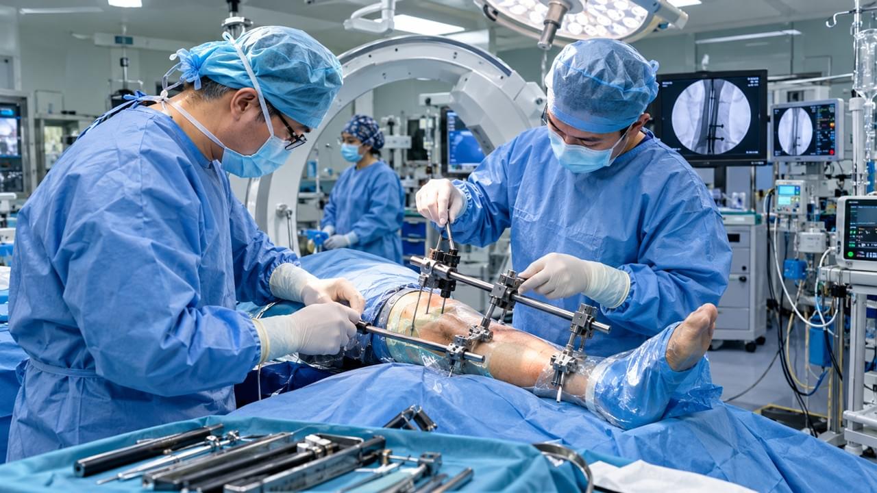
This article is part of our comprehensive guide: External Fixator Solutions: Types, Clinical Applications & Bone Fracture Recovery
TL;DR: Open (compound) fractures — where broken bone communicates with the outside world through a wound — are true orthopedic emergencies. 🚨 External fixators are the first-choice stabilization for severe open fractures (Gustilo Type III) because they deliver rapid bone stability without planting metal in contaminated tissue, keep the wound fully accessible, and enable definitive reconstruction once the soft tissue is ready. This guide covers the Gustilo-Anderson classification, antibiotic timing, debridement principles, fixator application, and staged reconstruction — six steps where minutes genuinely change outcomes.
⚠️ What Makes Open Fractures So Dangerous
Definition — Open Fracture: a fracture whose hematoma communicates with the external environment through a wound in the overlying skin. That wound might be a tiny puncture where a bone fragment speared through from the inside — or a gaping defect from high-energy trauma.
Why the clock starts the moment the injury happens:
- 🦠 Infection risk: the fracture site is contaminated with environmental pathogens. Left unmanaged, deep infection (osteomyelitis) rates exceed 30–50% in severe injuries
- 🩸 Soft tissue compromise: devitalized muscle, periosteum, and skin strip the fracture of its healing capacity
- 🫀 Vascular injury: high-energy patterns may involve arterial damage needing emergency revascularization
- 🚑 Systemic shock: femur and pelvis fractures cause major blood loss and hypovolemic shock
Open fractures make up just 2–4% of all fractures yet carry disproportionate morbidity. In low-resource settings, inadequate open fracture management remains a leading cause of preventable amputation and chronic osteomyelitis.
⭐ The one-paragraph version for the busy resident: resuscitate first, antibiotics within 1–3 hours, photograph and culture the wound without probing it, debride in the OR with generous low-pressure irrigation, stabilize severe injuries with a spanning external fixator, cover the wound early, and convert to definitive fixation only when the soft tissue says yes. Everything else in this article is detail around that skeleton — but the detail is where limbs are saved or lost.
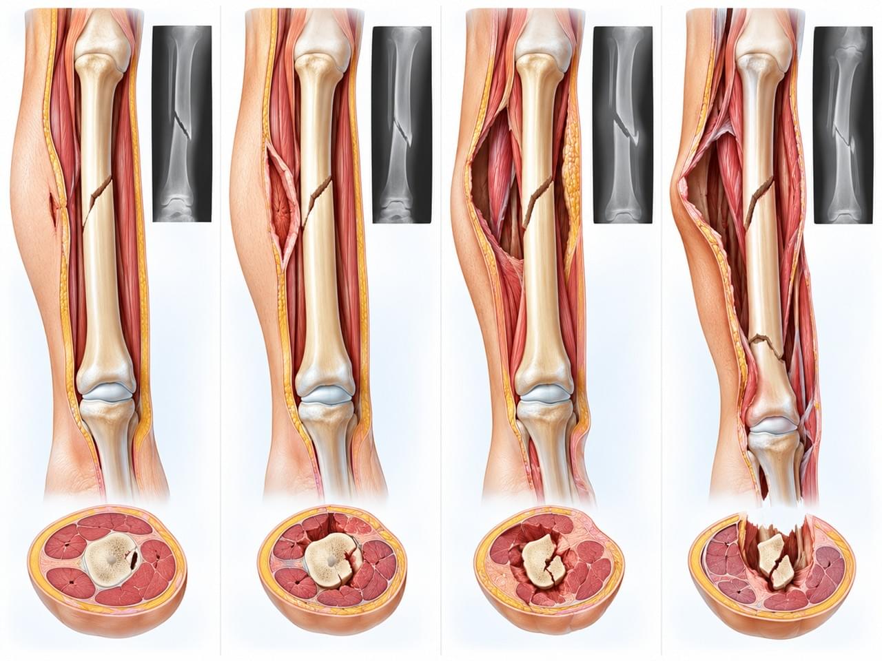
📊 Gustilo-Anderson Classification: The Universal Severity Language
The Gustilo-Anderson classification (1976, revised 1984) remains the international standard for grading open fracture severity — and for good reason. It predicts infection risk and drives the stabilization decision:
| Grade | Description | Infection Risk | Recommended Stabilization |
|---|---|---|---|
| Type I | Clean wound < 1 cm; simple pattern; minimal soft tissue damage | ~1–2% | Internal or external fixation equally appropriate |
| Type II | Wound 1–10 cm; moderate soft tissue damage; simple fracture | ~5–10% | Either method with good wound care |
| Type IIIA | Wound > 10 cm; extensive soft tissue injury; adequate bone coverage possible | ~10–20% | External fixation preferred |
| Type IIIB | Extensive periosteal stripping; coverage not possible; requires flap | ~20–50% | External fixation mandatory; staged reconstruction |
| Type IIIC | Any Type III with arterial injury requiring repair | >50% | External fixation + vascular surgery; amputation in select cases |
Full classification background is available from the AO Foundation (rel=”noopener noreferrer”, target=”_blank”). When in doubt between grades, chart the higher one — severity is consistently underestimated on first look.
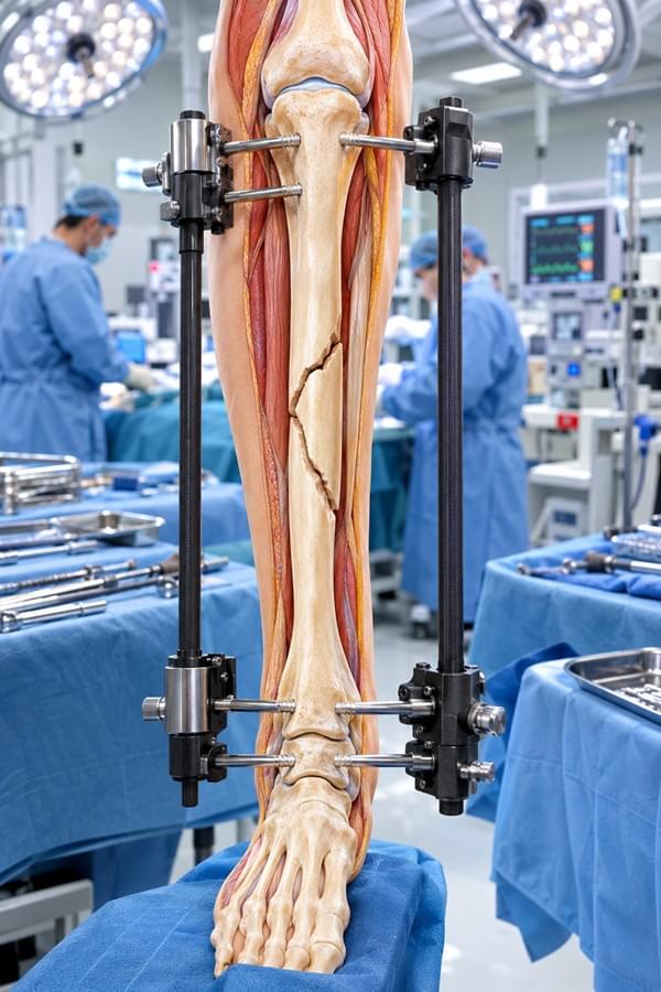
🚨 Emergency Management Protocol: 6 Time-Critical Steps
Step 1: Why Emergency Open Fracture Management Starts With Resuscitation
High-energy open fractures rarely travel alone. The primary survey (ABCDE — Airway, Breathing, Circulation, Disability, Exposure) takes absolute priority, and hemorrhage control with direct pressure precedes any fracture work. Sound trauma care starts with a patient well enough to survive the operating room. ⛑️
Step 2: Wound Assessment & Open Fracture First Aid
Good open fracture first aid — in the ER and at the scene — follows a disciplined sequence:
- 💧 Remove gross contamination (soil, debris) with saline irrigation
- 📸 Photograph the wound before any cleaning — medicolegal documentation matters
- 🧫 Take wound swabs for culture to guide definitive antibiotic therapy
- 🚫 Do NOT probe the wound extensively — it worsens contamination
- 🫁 Assess neurovascular status distal to the injury: pulses, capillary refill, sensation
Cover the wound with saline-moistened sterile dressings, splint the limb, and get imaging. Nothing more invasive before the OR.
Step 3: Antibiotic Timing — The Hour That Decides Open Fracture Management Outcomes
Antibiotic prophylaxis means systemic antibiotics given before bacterial colonization takes hold — prevention, not treatment. ⏱️ Administration within 1–3 hours of injury is strongly associated with reduced infection rates, and every hour of delay raises the risk. Of all the time-sensitive interventions in this protocol, this one shows the steepest penalty for waiting.
| Injury Grade | Recommended Antibiotic | Duration |
|---|---|---|
| Type I/II | Cephalosporin (1st gen, e.g., cefazolin 1g IV) | Until wound closure or 24 hrs |
| Type III | Cephalosporin + aminoglycoside (cefazolin + gentamicin) | 72 hrs post-injury max |
| Farm/soil contamination | Add metronidazole (anaerobic coverage) | 72 hrs |
| Tetanus prophylaxis | Per vaccination status | Single dose ± immunoglobulin |
(Protocols per EAST and AAOS open fracture guidelines)
Step 4: Irrigation & Debridement — The Cornerstone
Performed in the OR under anesthesia, I&D is where open fracture management is actually won or lost. 🧼
Irrigation: copious low-pressure saline — 3 liters minimum for Type I/II, 6–9 liters for Type III. Skip high-pressure pulsatile lavage: it damages periosteum and drives bacteria deeper into bone.
Debridement: systematic excision of all devitalized tissue —
- Skin: excise frank necrosis, preserve viable skin wherever possible
- Muscle: apply the 4 Cs — Color, Consistency, Contractility, Circulation; excise muscle failing 2+ criteria
- Bone: remove free contaminated fragments; keep periosteal-attached ones (they have blood supply)
Plan a repeat I&D at 48–72 hours — and repeat until the wound is genuinely clean. Single-visit debridement of a Type III injury is a gamble you rarely win.
💡 A hard-won field lesson: the most common debridement error is being too conservative with muscle. Necrotic muscle left in situ is a perfect culture medium — anaerobes thrive in it even after antibiotics. When the 4 Cs leave you uncertain about a muscle group, err toward excision; a functional deficit can be reconstructed later, but an abscess at the fracture site undoes everything else you’ve done.
Step 5: External Fixator Application in Open Fracture Management
Following I&D, skeletal stabilization is achieved with an external fixator. For Type III injuries, it is the default — and the reasoning is practical:
- 📍 Pins sit far from the contaminated fracture zone (spanning fixation)
- 🚫 No foreign metal in the infected or at-risk wound bed
- 🏗️ The frame provides rigidity enough for soft tissue healing and reconstruction
- 🩹 The wound stays fully accessible for repeat debridement and flap coverage
But why not just plate or nail it? Because metal implanted into a contaminated wound becomes a foreign-body surface bacteria love. Organisms adhere to implant surfaces and build biofilm — a protected state nearly impossible to eradicate without removing the implant. External fixation keeps hardware in clean tissue zones, away from the battlefield. This is the core logic of damage control orthopedics: stabilize fast, stay out of the wound, reconstruct later.
Application principles:
- Place pins in healthy cortical bone at least 5 cm from the wound edge
- Use spanning configurations: a bar bridging the fracture between proximal and distal pin blocks
- Ensure adequate rigidity so wound care doesn’t destabilize the construct
Technique details live in our minimally invasive external fixator insertion guide, and pin placement is faster and more precise with the right orthopedic power tools.
What about open femur fractures? A bilateral Type A configuration (two proximal and two distal pins, two parallel bars) or a “damage control” spanning fixator from iliac crest to proximal tibia provides adequate stability for resuscitation and soft tissue care. Plan early conversion to intramedullary nailing within 7–14 days once the patient is physiologically stable.
Step 6: Wound Coverage & Staged Reconstruction
- 🕐 Early coverage (within 72 hours): the best outcomes come from early definitive wound closure
- 🔬 Coverage options: primary delayed closure, split-thickness skin graft (STSG), local flap, or free microvascular flap for large defects
- 🔄 Conversion to internal fixation: once the wound is clean and covered — typically 7–14 days — the frame may be converted to definitive fixation
The choice between graft and flap is a soft tissue decision, not a bone one: STSG suits well-vascularized muscle beds, while exposed bone, tendon, or hardware demands flap coverage — local where possible, free microvascular for large defects. Getting plastic surgery involved early (ideally before the first debridement) saves days of sequential guesswork later. 🤝
When Does Open Fracture Management Turn Definitive?
Safe conversion timing is wound-driven, not calendar-driven. The wound must be clean, surgically closed or covered, and showing zero signs of infection — with all planned debridements complete. Premature conversion into an incompletely healed wound re-introduces exactly the infection risk you spent the first week fighting. Patience here is a surgical skill. 🧘
🌍 Equip Your Trauma Unit with Fixators Built for Emergencies
Vsun Medical’s external fixator range — from spanning trauma frames to mini digital fixators — is manufactured under GMP standards with ISO certification and a 2-year warranty, ready for global emergency supply. 📦
Browse the full external fixators product range or request trauma fixator specifications directly →. For how our manufacturing quality standards translate into field reliability, see quality standards in medical manufacturing.
GMP Compliant · ISO Certified · 2-Year Warranty · Global Emergency Supply Capability
Additional protocols are available from EAST .
 English
 Arabic
 German
 French
 Italian
 Spanish
 Russian
 Japanese
 Korean
 Turkish
 Portuguese