
📘 This article is part of our comprehensive guide: External Fixator Solutions: Types, Clinical Applications & Bone Fracture Recovery
TL;DR: Finger and metacarpal fractures are the most common upper extremity injuries in working-age adults, and they demand precision that casting alone cannot deliver. A mini external fixator maintains fracture reduction, allows wound management in open cases, and unlocks early digital rehabilitation — capabilities that translate into better functional outcomes. This guide covers epidemiology, fracture classification, mini fixator design and mechanics, surgical technique, rehabilitation phases, and how a mini fixator stacks up against K-wires, plates, and casting.
🩹 Hand Fractures: The Epidemiology & Clinical Challenge
Hand fractures — phalangeal (proximal, middle, distal) and metacarpal — account for roughly 10% of all fractures and are the most common fracture type requiring emergency department management in working-age adults.
Common mechanisms:
- 🏭 Crush injury — industrial/occupational
- 🏀 Sports trauma — ball sports, contact sports
- 🦴 Fall on outstretched hand — typical FOOSH pattern
- 🚗 Direct impact — motor vehicle accident
Clinical challenge: the hand is a precision instrument where even 5–10 degrees of rotational malreduction or articular incongruity translates into significant functional impairment. “Acceptable” fracture position in long bone surgery is simply not acceptable in hand surgery. Precision matters acutely.
Fracture Patterns That Need Surgical Fixation
Stable (conservative treatment appropriate):
- Non-displaced or minimally displaced (<2 mm) transverse fractures
- Spiral fractures with <5 mm shortening and no rotation
- Isolated distal phalanx tuft fractures
Unstable (surgical fixation indicated):
- 🦴 Displaced articular fractures — intra-articular fragments >2 mm displacement
- 🔄 Rotational deformities — any malalignment causing digital scissoring (fingers cross on flexion)
- 🩸 Open fractures — contaminated wounds preclude plate/screw internal fixation
- 💥 Comminuted fractures — multiple fragments making conventional fixation difficult
- ⚠️ Fracture-dislocations — combined joint dislocation with fracture (e.g., PIP joint)
- 🩹 Skin/soft tissue compromise — crush injuries with necrotic skin over the fracture site
🔧 Type A Design & Mechanics
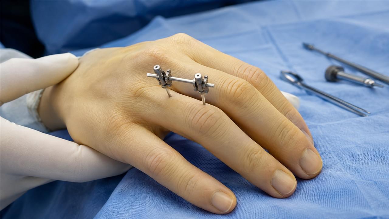
Type A is a linear (unilateral) external fixation system using miniaturized half-pins (1.0–2.5 mm diameter) inserted perpendicular to the long axis of the phalanx or metacarpal, connected by a compact bar and clamp system external to the skin. Compared to standard external fixation, the entire construct weighs 15–40 g — light enough to live on a finger for weeks.
Mini External Fixator Component Specifications
| Component | Specification |
|---|---|
| Half-pin diameter | 1.0 mm (distal phalanx) to 2.0 mm (proximal phalanx, metacarpal) |
| Pin material | Stainless steel or titanium alloy |
| Connecting bar | 3–4 mm diameter, carbon fiber or aluminum alloy |
| Bar length | 40–80 mm for digital applications |
| Clamp design | Simple pin-to-bar clamp; minimal bulk for digital application |
| Total weight | 15–40 g depending on configuration |
Biomechanical Advantages of the Mini External Fixator
- 🦴 Ligamentotaxis — the external frame maintains distraction across the fracture, using ligament and capsule tension to hold comminuted fragments in anatomical position; particularly valuable for articular fractures where individual fragment fixation is impossible
- 🔄 Three-dimensional alignment control — independent adjustment of pin angles and bar connection allows control of angular, rotational, and length deformity
- ⚡ Dynamic loading option — frame can be dynamized at callus formation stage to stimulate bone healing
🏥 Surgical Technique: Applying the Fixator
Step-by-Step Surgical Workflow
Equipment:
- 🔋 Battery-powered mini drill with 1.0–2.0 mm micro-drill bits
- 🧰 Type A fixator component set
- 📷 Fluoroscopy C-arm
- 🩸 Tourniquet (upper arm or digital)
Technique:
- Tourniquet inflation for bloodless field
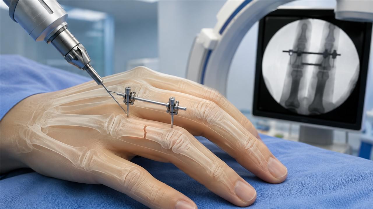
- Closed fracture reduction under fluoroscopy — traction, flexion, direct pressure
- Proximal pin placement — 1.5–2.0 mm half-pin through a 2–3 mm stab incision proximal to the fracture, avoiding extensor tendon and digital nerve
- Connecting bar application — bar and proximal clamp applied loosely
- Reduction maintenance — assistant holds reduction while distal pin is placed through second stab incision
- Frame locking — clamps tightened with fracture reduced, confirmed on fluoroscopy in two planes
- Wound closure — stab incisions closed with single suture; sterile pin site dressings
Surgical time: typically 20–35 minutes for a straightforward phalangeal fracture, significantly faster than open reduction and internal fixation (ORIF). The techniques pair well with our broader work on minimally invasive fixation — see Minimally Invasive External Fixator Insertion for the principles that apply across body sites.
Why the Mini External Fixator Wins for Hand Fractures
For finger fracture fixation and metacarpal fracture treatment, the device earns its place by doing what casting and K-wires struggle with: holding rotation, managing open wounds, and letting the patient move.
Mini fixator vs. K-wire fixation — what is the difference? K-wires are smooth percutaneous wires that may not provide complete rotational control and typically require removal under anesthesia. This device provides three-dimensional control (including rotation), allows wound management in open fractures, enables early digital rehabilitation while in place, and removes in clinic under local anesthesia. For complex, unstable, or open digital fractures, the difference is functional.
🩹 Rehabilitation: The Real Advantage of Early Mobilization
5 Phases of Mini External Fixator Recovery
| Timeframe | Rehabilitation Activities |
|---|---|
| 🧴 Days 1–3 post-op | Pin site care education; digital elevation; buddy taping if needed |
| 👐 Day 3–7 | Gentle active digital flexion/extension exercises within pain tolerance |
| 🤸 Weeks 2–4 | Progressive digital mobilization; commence hand therapy |
| ✅ Weeks 4–6 | Frame removal when radiographic healing confirmed; immediate unrestricted mobilization |
| 💪 Weeks 6–12 | Strengthening and functional rehabilitation |
How long does the fixator stay in place? For most phalangeal and metacarpal fractures, removal happens at 4–6 weeks when radiographic callus formation is confirmed. Articular fractures may require 6–8 weeks. Removal is performed in clinic under digital block local anesthesia and is well tolerated as an outpatient procedure.
Can Patients Use Their Hand Normally?
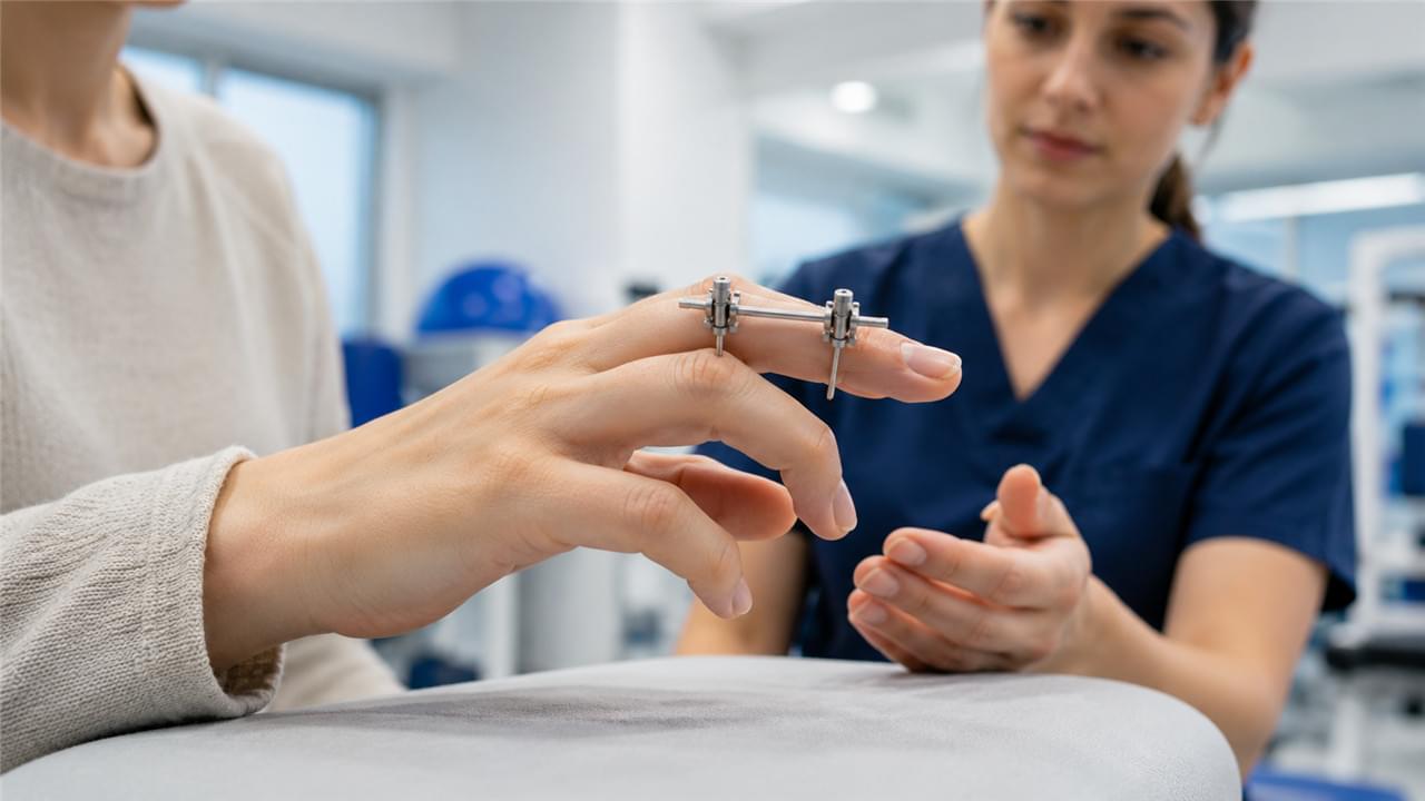
Patients can perform gentle daily activities (writing, computer use) and begin progressive digital rehabilitation while the mini fixator is in place, subject to surgeon guidance. Heavy lifting, grip activities, and contact sports are restricted until fracture union is confirmed and the frame is removed. Early mobilization during external fixation prevents the articular cartilage deterioration, synovial adhesions, and flexor tendon binding that are the primary complications of prolonged casting in hand injuries — and that is the single biggest reason hand surgery orthopedics has shifted toward dynamic fixation over the past two decades.
📊 How the Mini Fixator Stacks Up Against Alternatives
| Treatment Method | Fracture Access | Early Motion | Open Fracture Suitability | Revision Rate |
|---|---|---|---|---|
| Casting/Splinting | None needed | Not permitted | Contraindicated for open | Low |
| K-wire fixation | Percutaneous | Limited | Possible | Moderate (pin migration) |
| Plate/screw ORIF | Open dissection | Yes | Contraindicated for contaminated | Moderate |
| Type A mini fixator | Percutaneous | Yes, early | Well-suited | Low |
For complex phalangeal fractures — open, articular, or comminuted presentations — the fixator’s combination of wound access and early motion capability delivers superior functional outcomes compared to every alternative in the table.
✅ Supply Your Hand Surgery Unit with Precision Mini Fixator Systems
Vsun Medical’s Type A fixators — precision-machined for digital and metacarpal applications — are manufactured under GMP standards with ISO certification and a 2-year warranty.
What pin sizes do we supply? Vsun Medical’s fixator kits include half-pins in 1.0 mm, 1.2 mm, 1.5 mm, and 2.0 mm diameters for digital and metacarpal applications, with corresponding mini bars and clamps. Full product catalog and technical specifications are available upon request. Our broader range of external fixators covers everything from hand surgery to long-bone trauma.
Need a battery drill and bits for the procedure? See our Orthopedic Power Tools: Complete Guide for matching drill specifications.
Clinical technique and outcomes should follow established guidelines — the AAOS hand fracture management clinical guidelines and AO Foundation hand surgery fixation techniques are the most widely referenced resources.
📋 Request Mini Fixator Catalog & Pricing → vsunmedical.com/contact
GMP Compliant · ISO Certified · 2-Year Warranty · Global Supply
 English
 Arabic
 German
 French
 Italian
 Spanish
 Russian
 Japanese
 Korean
 Turkish
 Portuguese